Cutaneous vasculitis for the Dermatology SCE: exam guide
Revise cutaneous vasculitis for the UK Dermatology SCE: palpable purpura, biopsy timing, investigations, systemic red flags and treatment principles.
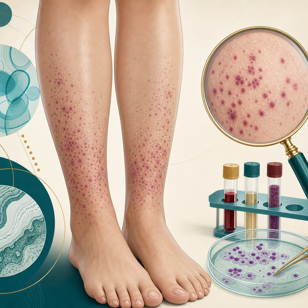
Cutaneous vasculitis questions test two skills at once. First, can you recognise inflammation of small vessels from the morphology? Second, can you decide whether it is confined to the skin or signals kidney, gut, nerve or lung disease? Palpable purpura gets you to the starting line. A safe investigation plan gets you the mark.
Recognise palpable purpura
The classic presentation of cutaneous small-vessel vasculitis is symmetrical palpable purpura on dependent areas, especially the lower legs. Purpura is red or purple because blood has escaped into the skin, so it does not blanch with pressure. It is palpable because inflammation and vessel-wall damage add oedema. New crops may appear over days, so lesions of different ages and colours can coexist.
- Typical small-vessel pattern: non-blanching palpable papules concentrated on the lower legs.
- Broader spectrum: petechiae, ecchymoses, urticarial plaques, haemorrhagic vesicles or bullae, necrosis and shallow ulceration can occur.
- Deeper vessel clue: livedo racemosa, painful subcutaneous nodules, punched-out ulcers or digital ischaemia should raise concern for medium-vessel disease.
- Common mimics: thrombocytopenia, coagulopathy, septic emboli, pigmented purpuric dermatosis and occlusive vasculopathy can produce purpura without primary vessel-wall inflammation.
The first safety decision: skin-limited or systemic?
Ask about organ symptoms before debating the cause. Cutaneous small-vessel vasculitis can be skin-limited, but the same eruption may accompany systemic vasculitis or another multisystem disease. Record blood pressure and look for oedema as well as examining the skin.
- Kidney: haematuria, proteinuria, reduced urine output, hypertension or worsening renal function.
- Gut: significant abdominal pain, gastrointestinal bleeding, vomiting or bowel ischaemia symptoms.
- Lung: dyspnoea, cough, chest pain or haemoptysis.
- Nerve: new foot drop, wrist drop, asymmetric sensory loss or other mononeuritis multiplex features.
- Systemic inflammation: fever, weight loss, marked fatigue, myalgia or severe arthralgia.
- ENT and eye clues: chronic sinus symptoms, epistaxis, hearing change, scleritis or other inflammatory eye symptoms can support ANCA-associated disease.
Organ red flags change the urgency and the team involved. Haemoptysis with renal abnormalities, rapidly progressive neuropathy, gastrointestinal bleeding or a severely unwell patient needs urgent hospital assessment. Do not label the eruption skin-limited until urinalysis and renal function have been checked.
Biopsy correctly: lesion choice and timing matter
Take two punch biopsies from fresh lesions when possible. A purpuric papule around 24 to 48 hours old is used for routine histology. A newer, partially blanching lesion is preferred for direct immunofluorescence because immunoglobulin deposits fade as a lesion ages. Histology of leukocytoclastic vasculitis shows a neutrophilic infiltrate around small vessels, nuclear dust from fragmented neutrophils, fibrinoid vessel-wall damage and extravasated red cells.
Baseline investigations that every candidate should know
The work-up has two purposes: find systemic involvement and look for a trigger. Start with urinalysis, blood pressure, full blood count, renal function, liver function and inflammatory markers. A chest radiograph is appropriate when there are respiratory symptoms or concern about systemic vasculitis. The clinical context then determines the second line.
- Urine microscopy and urine protein quantification if dipstick testing shows blood or protein.
- ANA and extractable nuclear antigens when connective tissue disease is plausible.
- ANCA when the pattern suggests granulomatosis with polyangiitis or microscopic polyangiitis.
- Complement levels and cryoglobulins for immune-complex or cryoglobulinaemic disease.
- Hepatitis B, hepatitis C and HIV testing when clinically indicated.
- Blood cultures and echocardiography when infective endocarditis is a possibility.
- Serum protein electrophoresis and age-appropriate malignancy assessment when the history points that way.
Causes: use the history before ordering the panel
Many episodes remain idiopathic, but a structured history often identifies the relevant category. Ask about infection, every prescription and non-prescription medicine, autoimmune symptoms and constitutional features. A drug eruption commonly begins days to weeks after exposure, but timing varies, and the suspected medicine should only be stopped or changed through appropriate clinical review.
- Drugs: antibiotics, non-steroidal anti-inflammatory drugs, thiazides, allopurinol and anticonvulsants are classic associations.
- Infections: streptococcal infection, hepatitis B or C, HIV and infective endocarditis are important exam associations.
- Autoimmune disease: systemic lupus erythematosus, rheumatoid arthritis, Sjogren syndrome and ANCA-associated vasculitis.
- Immune-complex disease: IgA vasculitis, cryoglobulinaemic vasculitis and hypocomplementaemic urticarial vasculitis.
- Other: inflammatory bowel disease, malignancy and monoclonal gammopathy enter the differential in the right context.
Named patterns the SCE may hide in the vignette
IgA vasculitis combines palpable purpura with arthralgia or arthritis, abdominal symptoms and renal involvement. Direct immunofluorescence shows vascular IgA deposition. It is commonest in children, but adult disease deserves careful renal surveillance because kidney involvement can be significant. Urticarial vasculitis produces wheals that persist for more than 24 hours, often burn or hurt, and leave bruising or pigmentation. Low complement and systemic features point towards hypocomplementaemic urticarial vasculitis.
ANCA-associated vasculitis is not diagnosed from the skin alone. Palpable purpura plus haematuria, pulmonary symptoms, neuropathy or destructive ENT disease should prompt urgent multispecialty assessment. Cutaneous polyarteritis nodosa affects medium vessels and is more likely to cause tender nodules, livedo and ulceration than fine palpable purpura.
Treatment principles
For a first episode that is confidently skin-limited, remove or treat an identified trigger and use rest, leg elevation, compression if appropriate, simple analgesia and skin care. Many episodes settle without systemic treatment. Persistent, recurrent, painful or ulcerating disease needs dermatology review. Colchicine, dapsone or a short corticosteroid course may be considered in selected cases, with monitoring and treatment tailored to comorbidity and cause.
Systemic vasculitis is different: therapy is directed by the organ-threatening diagnosis and usually requires rheumatology, renal, respiratory or other specialist involvement. Never let a topical treatment plan distract from abnormal urine, pulmonary haemorrhage, neuropathy or gastrointestinal disease.
The SCE vasculitis algorithm
- Confirm the morphology: palpable, non-blanching purpura on dependent sites.
- Search immediately for kidney, gut, lung, nerve and systemic red flags.
- Check blood pressure, urinalysis, full blood count and renal function.
- Biopsy a fresh purpuric papule for histology and a newer lesion for direct immunofluorescence.
- Use targeted serology and infection tests guided by the history and examination.
- Treat the trigger and skin symptoms only after dangerous systemic involvement has been addressed.
Connect this framework to cutaneous drug reactions, dermatological emergencies and skin of colour. Purpura may look less vividly red on richly pigmented skin, so palpation and attention to violaceous or brown colour change become especially important.
Put the sequence into practice with Dermatology SCE questions: morphology, organ screen, biopsy, targeted cause, then treatment.
Sources & further reading
- Cutaneous small vessel vasculitis - DermNet
- Cutaneous vasculitis: an algorithmic approach to diagnosis - Frontiers in Medicine
- A rheumatology perspective on cutaneous vasculitis - Clinical Medicine
- Update on vasculitis: an overview and dermatological clues - Anais Brasileiros de Dermatologia
- Management recommendations for ANCA-associated vasculitis - British Society for Rheumatology
For educational use only. Check current official guidance.
Start 25 free questions