Dermatopathology in the Dermatology SCE: how to read histology under pressure
A practical approach to dermatopathology spot-diagnosis questions in the Dermatology SCE: the reaction patterns examiners reuse, the classic give-away clues, and how to build recognition fast.
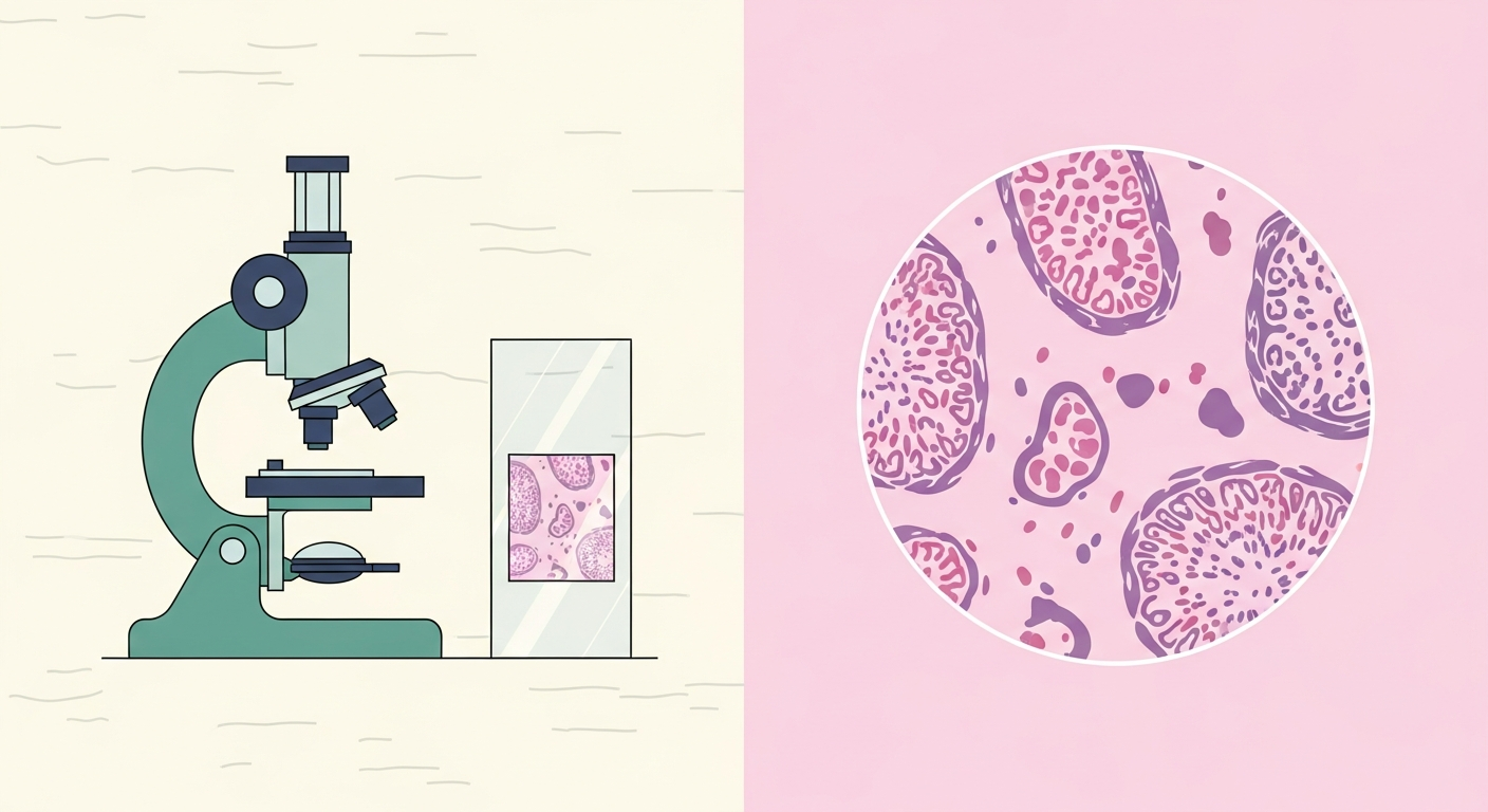
Dermatopathology is the part of the SCE that candidates most often dread and most often underprepare. Yet histology questions are some of the most predictable marks in the paper, because the exam reuses a defined set of reaction patterns and a familiar list of give-away clues. Approach them systematically and they become reliable points.
Work by reaction pattern first
- Spongiotic (intercellular oedema): the eczema group.
- Psoriasiform (regular acanthosis): psoriasis and its mimics.
- Lichenoid and interface (basal damage at the dermo-epidermal junction): lichen planus, lupus, lichenoid drug eruptions.
- Vesiculobullous (blisters, sub-epidermal or intra-epidermal): the immunobullous group.
- Granulomatous: sarcoid, granuloma annulare, necrobiosis lipoidica, infection.
- Vasculitic, depositional, panniculitic and neoplastic complete the common patterns.
The give-away clues examiners reuse
- Sawtooth rete ridges, a band of lymphocytes and Civatte bodies: lichen planus.
- Sub-epidermal blister with eosinophils: bullous pemphigoid.
- Intra-epidermal acantholysis, a row of tombstones: pemphigus vulgaris.
- Festooning of dermal papillae and caterpillar bodies: porphyria cutanea tarda.
- Palisading granuloma with central mucin: granuloma annulare.
- Naked dermal granulomas: sarcoidosis.
- Storiform spindle cells with a grenz zone, CD34 positive: dermatofibrosarcoma protuberans, as opposed to a CD34-negative dermatofibroma.

Special stains and immunohistochemistry worth knowing
- PAS: fungi and the basement membrane.
- Ziehl-Neelsen: acid-fast bacilli.
- Congo red: amyloid, with apple-green birefringence under polarised light.
- Perls Prussian blue: iron.
- Melan-A, SOX10 and S100: melanocytic lesions.
- CD34: positive in dermatofibrosarcoma protuberans, negative in dermatofibroma.
Direct immunofluorescence patterns
- Linear IgG and C3 along the basement membrane: bullous pemphigoid.
- Intercellular IgG, a chicken-wire or net-like pattern: pemphigus.
- Granular IgA in the dermal papillae: dermatitis herpetiformis.
- IgA along the basement membrane: linear IgA disease.
How to build recognition
- Drill image questions rather than reading pathology passively. Recognition is a visual skill.
- Always start from the reaction pattern, then apply the specific clue. The algorithm protects you when a slide is unfamiliar.
- Use spaced repetition with image cards that pair each pattern and clue with its diagnosis.
Sources & further reading
- DermNet - pathology of skin conditions
- PathologyOutlines - skin nontumour and tumour
- British Association of Dermatologists - clinical guidelines
For educational use only. Check current official guidance.
Start 25 free questions