Immunobullous disease in the SCE: pemphigus, pemphigoid and the blistering traps
High-yield immunobullous revision for the UK Dermatology SCE: pemphigus vs pemphigoid, immunofluorescence patterns, Nikolsky sign, dermatitis herpetiformis and management ladders.
Examiners love blistering diseases because they test a clean chain of reasoning: clinical pattern, level of the split, immunofluorescence, then management. The good news is that a small number of facts unlocks nearly every question. This guide gives you those facts.
First decision: where does the skin split?
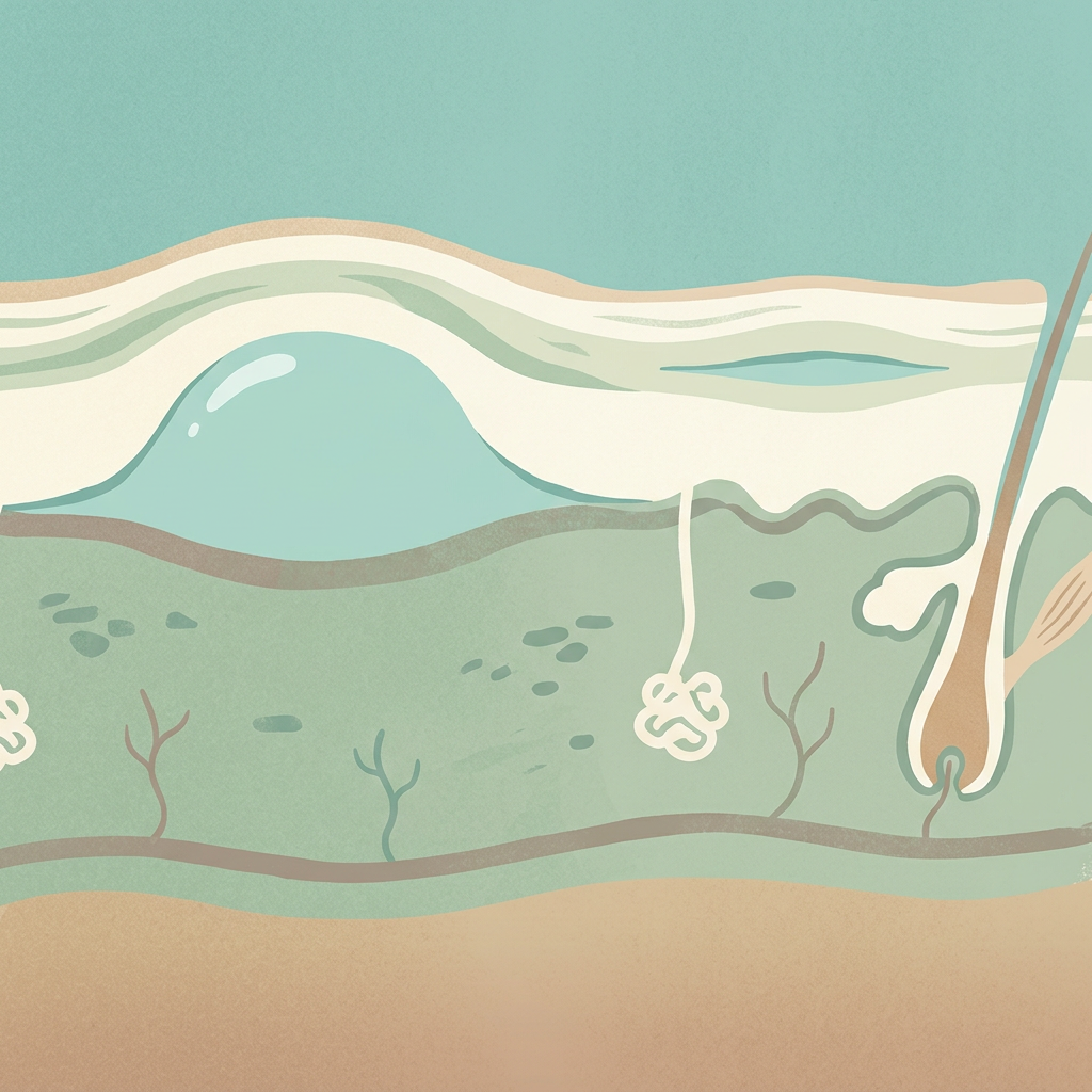
Everything follows from the level of cleavage. An intraepidermal split produces flaccid, fragile bullae that rupture almost immediately, leaving painful erosions, that is the pemphigus group. A subepidermal split produces tense, durable bullae that can sit intact for days, that is the pemphigoid group. Read the description of the blisters first and the diagnosis is usually halfway done.
- Pemphigus vulgaris, antibodies against desmoglein 3 (plus or minus desmoglein 1); painful mucosal erosions usually come first (mouth especially), then flaccid bullae; Nikolsky sign positive; suprabasal acantholysis on histology.
- Pemphigus foliaceus, antibodies against desmoglein 1; superficial crusted erosions in a seborrhoeic distribution (face, scalp, upper trunk); no mucosal involvement; subcorneal split.
- Bullous pemphigoid, antibodies against BP180 and BP230 hemidesmosomal antigens; elderly patients; tense bullae on flexures and lower abdomen, often after an itchy pre-bullous phase; mucosa usually spared; Nikolsky negative.
- Dermatitis herpetiformis, intensely itchy grouped vesicles on elbows, knees, scalp and buttocks; the cutaneous marker of coeliac disease.
Immunofluorescence: the vocabulary the exam uses
Direct immunofluorescence is taken from perilesional skin. Intercellular chicken-wire IgG within the epidermis means pemphigus. Linear IgG and C3 along the basement membrane zone means bullous pemphigoid. Granular IgA in the dermal papillae means dermatitis herpetiformis. Salt-split skin refines the subepidermal diagnoses: staining on the epidermal side (the roof) points to BP180 disease, while staining on the dermal side (the floor) points to epidermolysis bullosa acquisita. Serology, anti-Dsg1, anti-Dsg3, anti-BP180, anti-BP230 ELISAs, confirms the diagnosis and tracks activity.
Dermatitis herpetiformis deserves its own line
Dermatitis herpetiformis is the skin expression of coeliac disease: granular IgA deposits, positive anti-tissue transglutaminase and endomysial antibodies, and often no bowel symptoms at all. Treat with dapsone, check G6PD first because of haemolysis risk, and remember methaemoglobinaemia, alongside a strict gluten-free diet, which is the disease-modifying answer. Do not let the word herpetiform push you towards herpes; the name describes the grouped morphology only.
Management ladders
Bullous pemphigoid: superpotent topical corticosteroid (clobetasol propionate) applied to the whole body is first line even in generalised disease, the trial evidence showed it beats oral prednisolone for safety. Doxycycline is a useful steroid-sparing option, with azathioprine, methotrexate or mycophenolate for difficult disease. Pemphigus vulgaris: rituximab plus prednisolone is now first line for moderate-to-severe disease, the Ritux 3 trial changed the exam answer, and papers written after it expect you to know this. Historically the answer was high-dose prednisolone with azathioprine.
Mucous membrane pemphigoid is the dangerous cousin: a scarring subepidermal disease where ocular involvement can blind. It needs systemic immunosuppression, dapsone for moderate disease, cyclophosphamide for severe or rapidly progressive ocular disease, plus ophthalmology involvement from the start. Never manage it like ordinary bullous pemphigoid.
The rest of the blister differential
- Linear IgA disease, linear IgA at the basement membrane; annular string-of-pearls blistering; vancomycin is the classic drug trigger; dapsone responsive.
- Epidermolysis bullosa acquisita, antibodies against type VII collagen; trauma-induced blisters that heal with scarring and milia.
- Pemphigoid gestationis, BP180 autoimmunity in pregnancy, often starting periumbilically in the second or third trimester; associated with fetal growth restriction and prematurity.
- Paraneoplastic pemphigus, severe, intractable mucosal disease with an underlying malignancy (lymphoma, Castleman disease); remember bronchiolitis obliterans as its feared complication.
How to revise blisters for the SCE
Build a two-column table, pemphigus versus pemphigoid, and add the immunofluorescence row until it is automatic. Then connect this topic to its neighbours: dermatological emergencies (SJS/TEN shares the positive Nikolsky sign) and cutaneous drug reactions (vancomycin and linear IgA disease).
Want to drill this properly? Practise immunobullous questions in the Derm SCE question bank, image-led cases with explanations referenced to the literature.
Sources & further reading
- Bullous pemphigoid - NICE Clinical Knowledge Summaries
- Pemphigus vulgaris - DermNet NZ
- Bullous pemphigoid - DermNet NZ
- Dermatitis herpetiformis - DermNet NZ
- First-line rituximab combined with short-term prednisone versus prednisone alone for pemphigus (Ritux 3) - PubMed
For educational use only. Check current official guidance.
Start 25 free questions