Hair loss for the Dermatology SCE: an alopecia guide
Revise hair loss for the UK Dermatology SCE: alopecia areata, telogen effluvium, scarring alopecia, investigations, treatment and exam traps.
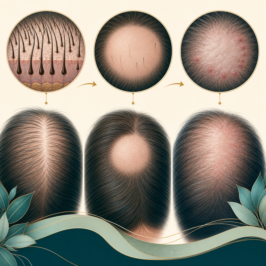
Hair loss is a gift to the well-organised candidate. The list of diagnoses looks long, but most vignettes can be solved with three decisions: is the loss focal or diffuse, are follicular openings still visible, and is there active inflammation? This guide turns those decisions into a repeatable approach for the Dermatology SCE.
First decide whether the alopecia is scarring
Look closely at the scalp rather than at the amount of hair. In non-scarring alopecia, follicular openings remain visible and regrowth is possible because the follicle is still present. In scarring alopecia, inflammation destroys the follicle and replaces it with fibrosis. The surface looks smooth and shiny, with absent follicular openings. This distinction matters more than almost any individual blood test because scarring alopecia needs early treatment to prevent permanent loss.
- Smooth focal patches with preserved openings: think alopecia areata. Exclamation-mark hairs, yellow dots and black dots support the diagnosis.
- Diffuse shedding with a normal-looking scalp: think telogen effluvium, especially around 3 months after illness, surgery, childbirth, marked weight loss, psychological stress or a medication change.
- Frontotemporal recession with eyebrow loss: think frontal fibrosing alopecia, a patterned form of lichen planopilaris.
- Perifollicular scale, erythema, pustules or crusting with lost openings: think an active primary scarring alopecia and biopsy the active edge.
- Scale, broken hairs and lymphadenopathy in a child: think tinea capitis rather than alopecia areata.
Alopecia areata: the classic non-scarring patch
Alopecia areata is an immune-mediated, non-scarring alopecia. The classic lesion is a sharply defined round or oval patch of smooth, normal-coloured scalp. Exclamation-mark hairs taper towards the scalp at the active margin. Nail pitting or rough nails can accompany the hair loss. Ophiasis describes a band around the occipital and temporal margins; alopecia totalis affects the whole scalp, while alopecia universalis affects scalp and body hair.
Assess the extent and activity, examine eyebrows, eyelashes, nails and the rest of the skin, and ask about autoimmune disease. Thyroid disease is an association, but a broad laboratory screen is not automatically required in every typical case. Investigations should be driven by the history, examination and differential. The diagnosis is usually clinical, supported by dermoscopy when needed.
Alopecia areata treatment in a UK question
Management depends on age, extent, duration and impact. Limited patch disease may be observed because spontaneous regrowth is possible, or treated with a potent topical corticosteroid. Intralesional corticosteroid is a useful specialist option for limited disease in adults. More extensive disease should prompt dermatology referral for options such as contact immunotherapy or systemic treatment. NICE recommends the oral JAK inhibitor ritlecitinib, within its marketing authorisation, for severe alopecia areata in people aged 12 years and over. NICE also allows deuruxolitinib within its marketing authorisation for severe disease in adults, with CYP2C9 genotyping before treatment. Treatment can induce regrowth but does not remove the tendency to relapse, so avoid promising a cure.
Telogen effluvium: connect the shedding to the timeline
Telogen effluvium is diffuse, non-scarring shedding caused when more follicles than usual enter the resting phase. The trigger commonly precedes the shedding by about 3 months, so ask backwards: febrile illness, major surgery, childbirth, crash dieting, iron deficiency, thyroid disease, severe stress or a new medicine may have been forgotten by the time the hair starts to fall. The scalp usually looks normal and the patient notices hair in the shower, on the pillow or in the brush rather than one bald patch.
A hair-pull test may be positive during active shedding. Check the whole scalp for pattern loss or inflammation and use targeted tests when the history suggests an underlying driver, commonly a full blood count, ferritin and thyroid function. Reassurance and correction of the trigger are the treatment. Shedding often lasts for 3 to 6 months, but visible density takes longer to recover because new hair grows slowly. Chronic or unexplained shedding deserves reassessment rather than repeated supplements without evidence of deficiency.
Scarring alopecia: recognise activity before the scar
Primary scarring alopecias are uncommon but disproportionately important because delay makes the loss irreversible. Symptoms such as itch, pain or burning, and signs such as perifollicular erythema, scale, pustules and crusting, identify activity. Smooth pale areas with no follicular openings show established damage. The treatment goal is to stop further loss, not to regrow follicles that have already been destroyed.
- Lichen planopilaris: irregular patches with perifollicular erythema and scale, often with itch or burning.
- Frontal fibrosing alopecia: a symmetrical band of frontotemporal recession, commonly with early eyebrow loss and perifollicular scale at the hairline.
- Discoid lupus erythematosus: erythematous scaly plaques with follicular plugging, dyspigmentation and atrophy.
- Central centrifugal cicatricial alopecia: begins at the crown and expands centrifugally; it is particularly important to recognise in women of African ancestry.
- Folliculitis decalvans: a neutrophilic pattern with pustules, crusting and tufted hairs.
If scarring alopecia is suspected, refer early and biopsy an active margin rather than the smooth centre of an old scar. The histology request should explain the suspected alopecia and biopsy site. Paired punch biopsies may be used for horizontal and vertical sections, with a further specimen for direct immunofluorescence when lupus or another immune-mediated process is in the differential. Treatment is diagnosis-specific and specialist-led, using anti-inflammatory or antimicrobial therapy to suppress activity.
The SCE hair loss algorithm
- Decide whether the loss is focal, patterned or diffuse.
- Look for follicular openings. Preserved means non-scarring; absent means scarring until proved otherwise.
- Look for activity: scale, erythema, pustules, crusting, broken hairs or a positive pull test.
- Use the timeline. Sudden diffuse loss about 3 months after a trigger strongly supports telogen effluvium.
- Order targeted investigations rather than a reflex panel, and biopsy the active edge when scarring is suspected.
- Choose the management aim: encourage regrowth in non-scarring disease, but halt progression in scarring disease.
Hair loss links several parts of the syllabus. Pair this guide with paediatric dermatology for tinea capitis, genodermatoses for inherited hair disorders, and dermatopathology for biopsy pattern recognition.
Ready to test the framework? Practise Dermatology SCE questions and force yourself to make the scarring versus non-scarring decision before reading the options.
Sources & further reading
- Alopecia areata - NICE Clinical Knowledge Summaries
- Ritlecitinib for treating severe alopecia areata in people 12 years and over (TA958) - NICE
- Deuruxolitinib for treating severe alopecia areata (TA1178) - NICE
- Alopecia areata - British Association of Dermatologists
- Telogen effluvium - British Association of Dermatologists
- Primary cicatricial alopecia: diagnosis and treatment - Canadian Medical Association Journal
For educational use only. Check current official guidance.
Start 25 free questions