Skin cancer in the Dermatology SCE: melanoma, BCC and SCC high-yield guide
High-yield skin cancer revision for the UK Dermatology SCE: melanoma Breslow thickness and staging, BCC subtypes, SCC risk factors, excision margins and the traps examiners set.
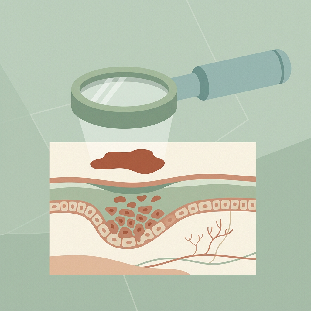
Skin cancer is the largest single clinical area in the Dermatology SCE, and it rewards structured revision more than almost anything else. Every year the paper tests the same core skills: recognise the tumour from a description or image, name the prognostic factors, and choose the right management. This guide covers exactly that core, nothing more, nothing less.
How the SCE tests skin cancer
Questions cluster around three moves. First, diagnosis: a pearly papule with telangiectasia, a changing mole, a crusted nodule on sun-damaged skin. Second, prognosis: Breslow thickness, ulceration, high-risk features. Third, management: excision margins, when to choose Mohs, when to refer for sentinel node biopsy or adjuvant therapy. Vignettes love an immunosuppressed transplant patient or an elderly patient with a lifetime of sun exposure, if you see either, raise your suspicion threshold immediately.
Melanoma: the highest-stakes topic
The UK 7-point checklist scores major features (change in size, change in shape, change in colour, 2 points each) and minor features (diameter 7 mm or more, inflammation, itch or altered sensation, oozing or crusting, 1 point each). A score of 3 or more warrants urgent referral. The ABCDE rule (asymmetry, border, colour, diameter, evolution) is the public-facing adjunct. Crucially, nodular melanoma often fails ABCDE, remember EFG instead: elevated, firm, growing.
Breslow thickness, measured in millimetres from the top of the granular layer to the deepest tumour cell, is the single most important prognostic factor, and ulceration upstages the tumour; Clark level has been abandoned. Under AJCC 8, T1 is 1.0 mm or less (T1b is 0.8–1.0 mm or any thinner tumour with ulceration). Sentinel lymph node biopsy is discussed from T1b upwards. For stage III and resectable stage IV disease, test for BRAF V600 mutations: adjuvant options are checkpoint immunotherapy (nivolumab or pembrolizumab) or, if BRAF-mutant, targeted therapy with dabrafenib plus trametinib.
Basal cell carcinoma
The commonest skin cancer: a pearly papule or nodule with surface telangiectasia and a rolled edge, usually on sun-exposed sites. Know the subtypes, nodular (commonest), superficial (younger patients, trunk, often multiple), morphoeic or infiltrative (scar-like, ill-defined, the worst for incomplete excision) and basosquamous. Risk factors: ultraviolet exposure, fair skin, immunosuppression, and Gorlin syndrome (autosomal dominant PTCH1 mutation, multiple early BCCs, jaw keratocysts, palmar pits, bifid ribs).
Management follows risk. Low-risk tumours: standard excision with a 4–5 mm margin, or curettage and cautery. Superficial BCC: imiquimod, 5-fluorouracil, photodynamic therapy or cryotherapy. High-risk tumours, morphoeic histology, recurrent lesions, and the facial H-zone around eyes, nose and ears, go to Mohs micrographic surgery. Locally advanced or metastatic BCC: vismodegib, a hedgehog pathway inhibitor (remember its signature adverse effects: teratogenicity, muscle spasms, dysgeusia, alopecia).
Cutaneous squamous cell carcinoma
A firm, keratotic, often crusted nodule that grows faster than a BCC and may ulcerate. The precursor continuum matters: actinic keratosis to Bowen's disease (SCC in situ, a well-defined scaly plaque) to invasive SCC. SCC arising in a chronic scar or ulcer is a Marjolin ulcer. Immunosuppression multiplies the risk dramatically, so the classic vignette is a renal transplant recipient developing multiple aggressive SCCs years after transplantation.
High-risk features to recite: diameter 2 cm or more, poor differentiation, perineural invasion, invasion beyond fat or deep, site on the lip or ear, and immunosuppression. Standard excision margins are about 4 mm for low-risk and 6–10 mm for high-risk tumours, and you always palpate the regional lymph nodes, unlike BCC, SCC metastasises. Radiotherapy is the adjuvant option, and cemiplimab (anti-PD-1) treats advanced disease.
The rare tumours that appear every year
- Merkel cell carcinoma, driven by Merkel cell polyomavirus; a rapidly growing red–violaceous nodule on sun-damaged elderly skin; aggressive, with early nodal spread.
- Dermatofibrosarcoma protuberans, a slow, indurated plaque, usually on the trunk; CD34 positive with a COL1A1–PDGFB fusion; locally aggressive, so wide excision or Mohs.
- Mycosis fungoides (cutaneous T-cell lymphoma), patches and plaques in a bathing-trunk distribution that may itch for years; Sezary syndrome is erythroderma with circulating Sezary cells and lymphadenopathy.
- Kaposi sarcoma, HHV-8; violaceous patches and nodules; classic form affects the legs of elderly Mediterranean or Eastern European men, with immunosuppression-related forms alongside.
How to revise skin cancer for the SCE
Work through image-rich questions until recognition is instant, then drill the numbers: margins, Breslow thresholds, high-risk features. Pair this guide with our posts on dermoscopy patterns, skin surgery and margins and genodermatoses (for Gorlin syndrome). Consolidation beats volume here.
Ready to test yourself? Start practising with the Derm SCE question bank, every question in the tumours section mirrors these exam patterns, with referenced explanations.
Sources & further reading
- Suspected cancer: recognition and referral (NG12) - NICE
- Melanoma - DermNet NZ
- Melanoma skin cancer statistics - Cancer Research UK
- Basal cell carcinoma - DermNet NZ
- Cutaneous squamous cell carcinoma - DermNet NZ
For educational use only. Check current official guidance.
Start 25 free questions